JEOL is industrial manufacturer of Electron Microscope and leader in this technology. has done an excellent study on the distinction between different asbestos fibers in Transmission Electron Microscopy. I would like to share with you this excellent publication.
good reading ………………………….. N.Benmeradi, R & D DeltaMicroscopies France
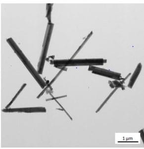
Transmission Electron Microscope (hereinafter, abbreviated to “TEM”) allows for not only observation of images of internal structure/morphology and of electron diffraction patterns, but also elemental analysis combined with Energy Dispersive X-ray Spectroscopy (hereinafter, abbreviated to “EDS”). Presented here are analysis results of Amosite, Crocidolite, Chrysotile, Anthophyllite and Tremolite/Actinolite. Those asbestos were analyzed by morphological observation of TEM images, electron diffraction and elemental analysis using EDS. Since various asbestos are enabled to be identified through differences in their fibrous shapes, electron diffraction patterns and constituent elements, TEM can distinguish asbestos only from one fiber.
Types of asbestos
| asbestos | Category | Name of mineral | Name of asbestos | Chemical formula | H | O | Si | Na | Mg | Ca | Fe |
|---|---|---|---|---|---|---|---|---|---|---|---|
| Serpentine group | chrysotile | Serpentine, Chrysotile | Mg3Si2O5(OH)4 | ○ | ○ | ○ | ○ | ||||
| Amphibole group | amosite | Amosite | (Fe2+, Mg)7Si8O22(OH)2 | ○ | ○ | ○ | ○ | ○ | |||
| crocidolite | Crocidolite | Na2(Fe2+, Mg)3Fe3+2Si8O22(OH)2 | ○ | ○ | ○ | ○ | ○ | ○ | |||
| anthophyllite#1 | Anthophyllite | (Mg, Fe2+)7Si8O22(OH)2 | ○ | ○ | ○ | ○ | ○ | ||||
| tremolite#1,2 | Tremolite | Ca2(Mg, Fe2+)5Si8O22(OH)2 | ○ | ○ | ○ | ○#2 | ○ | ○#2 | |||
| actinolite#1,2 | Actinolite | Ca2(Fe2+ ,Mg)5Si8O22(OH)2 | ○ | ○ | ○ | ○#2 | ○ | ○#2 |
Chrysotile Mg3Si2O5(OH)4


Amosite (Fe2+, Mg)7Si8O22(OH)2

 (The spectral peaks of C and Cu are generated from the elements of the support film and the grid.)
Crocidolite Na2(Fe2+, Mg)3Fe3+2Si8O22(OH)2

 (The spectral peaks of C and Cu are generated from the elements of the support film and the grid.)
anthophyllite (Mg, Fe2+)7Si8O22(OH)2


 (The spectral peaks of C and Cu are generated from the elements of the support film and the grid.)
Jeol Differents Asbestos Fibers Electron Microscopy
Tremolite / Actinolite Ca2(Mg, Fe2+)5Si8O22(OH)2 / Ca2(Fe2+ ,Mg)5Si8O22(OH)2


 (The spectral peaks of C and Cu are generated from the elements of the support film and the grid.) The mineral types of Tremolite and Actinolite are different depending on the content difference in Mg and Fe.


