Description
NIOPROBE
The Nioprobe device is used to determine the shape at the very apex of the tip probe for microscopy.
The Problem
The physical probe used in AFM imaging is not ideally sharp. As a consequence, an AFM image does not reflect the true sample topography impartially, but rather represents the interaction of the tip with the sample surface. There is no avoiding this imperfection, which sets real limits on what may be validly inferred from an AFM image.
Whether one is engaged in detailed, quantitative metrology or is simply using AFM images as a interpretive aid, it is imperative be able to assess these limits. The key here is to possess a reliable estimate of the sharpness of the tip apex. Reverse imaging of the probe is the most convenient means of obtaining the effective radius of the probe. For this purpose, the ideal characterization sample would consist of small, stiff, spiked features.
The Practical Answer is NioProbe
Consider the following advantages of NioProbe:
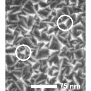
– The surface structure of the NioProbe film is densely populated by tiny peaks. This makes the film very suitable for the small piezo movements characteristic of precision AFM work.
– Feature peaks exhibit imaging radii of less than 5 nm, as sharp as anything else available. This permits one to obtain the accurate apex radius desired for medium- to small- scale work (such as biomolecular imaging).
– The random orientation of the NioProbe features are suitable for applying blind tip reconstruction methods.
– The sample is resistant to the duress of contact mode scanning.
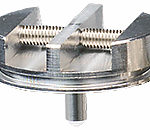
– The film is supplied on a silicon chip, ready to be placed in your AFM. Instructions are provided to allow easy determination of the apex radius. If stored in a clean, dry place, the sample can provide years of service.

Reviews
There are no reviews yet.