Description
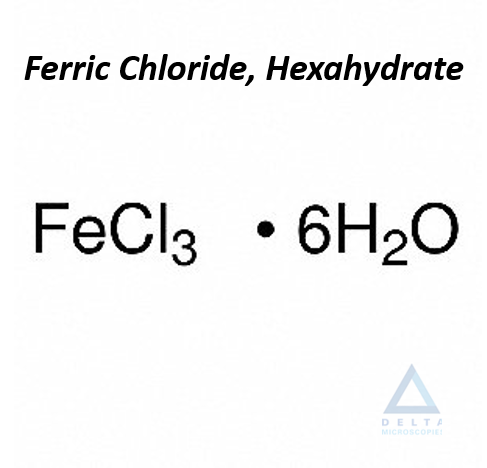
FeCl3·6H2O F.W. 270.30 CAS #13478-10-9
| Assay | 97.0-102% |
| Insoluble Matters. | 0.01% |
| Nitrate (about 0.001%) | To Pass Test |
| Phosphorous Compound | 0.01% |
| Sulfate | 0.01% |
| Arsenic | 0.002% |
| Copper | 0.003% |
| Zinc | 0.003% |
References for use: Membrane stains – Gasic, G., Berwick et al (1963). Hale stain for sialic acid containing mucins. J. Cell Biol., 19:223 – Gasic, G., Berwick, et al (1968). Positive and Negative colloidal iron as cell surface electron stains. Lab. Invest., 18:63 – Blanquet, P.R. and Loiez, A. (1974). Colloidal iron used at pHs lower than 1 as electron stain for surface proteins. J. Histochem. Cytochem., 22:368 – Matukafs, V.J., Panner et al (1967). Studies on ultrastructural identification and distribution of protein-polysaccharides in cartilage matrix. J. Cell Biol., 32:365 – Benedetti, E.L. and Emmelot, P. (1967). Studies on plasma membranes IV. The Ultrastructural localization and content of sialic acid in plasma membranes isolated from rat liver and hepatoma. J. Cell Sci., 2:499 – Rowley, J.R. (1971). Resolution of channels in the exine by translocation of colloidal iron. Pro. 29th Ann. EMSA Meet., p.352. Claitor’s Pub. Division, Baton Rouge, LA – Nicolson, G.L. (1973). Anionic sites of human erythrocyte membranes.I. Effects of trypsin, phospholipase C, and pH on the topography of bound positively charged colloidal particles. J. Cell Biol., 57:373 – Hendy, R. (1971). Electron microscopy of lipofucsin pigment stained by the Schmorl and Fontana technique. Histochemie, 26:311.
Storage : RT



Avis
Il n’y a pas encore d’avis.