Description
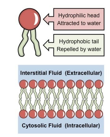
A new histochemical method is described for the polychromatic staining and identification of phospholipids and sulpholipids in tissue sections. It is an adaptation of a chromatographic method described previously (Kennedy & Collier, 1962). Sections are stained in a mixture of Diamond Sky Blue B, Diamond Cyanine R and formalin, followed by differentiation in 2-morpholinoethanol and bromine water, counterstaining in Cresyl Fast Violet and decolorization in lactic acid.
The interpretation of the final colours in the test tissues was checked by extraction and chromatography of their lipid content.
The staining of diverse animal and plant tissues is described and illustrated in colour.
Stain results:
| Phospholipids: | Blue | |
| Nuclei and Nucleoli: | Red | |
| Early lipofuchsin, eosinophil granules, keratin, keratohyalin, human elastic tissues: |
Blue to Purple |
References:
Pearse, A. G.E., J. Pathl. Bact., 70:554-557, 1995
Luna, L.G. (ed), Manual of Histologic Staining Methods of the Armed Forces Institute of Pathology, 3rd Ed., McGraw-Hill, c. 1968, p. 148.




Avis
Il n’y a pas encore d’avis.