Technical Blog
You Find :
- Definitions of some technicals terms for more precision..
- Tips, advice, recommendations, protocols and methods.
- Storie of microscopes and microscopists, more news to our microscopy planet.

Definitions
For more precision
Methods
always the same passion
Microscopy Land
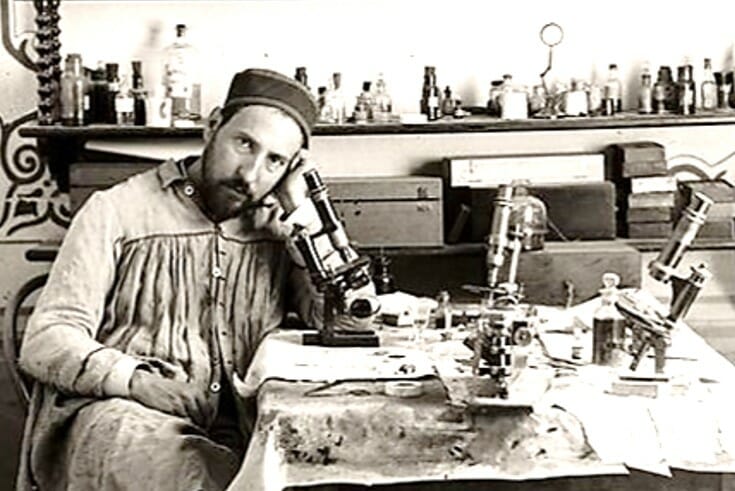
history and stories
In this blog by keywords or Tag, you will find various information which can help you during your investigations in microscopy.
Definitions :
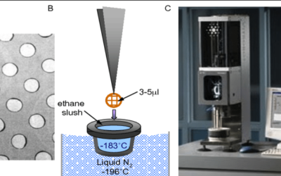
QUANTIFOIL® Holey Carbon Films
application : Cryo-EM-sample-freezing-A-Portion-of-a-Quantifoil-TM-holey-gridQUANTIFOIL® is a perforated support foil with a pre-defined hole size, shape and arrangement. We offer a variety of QUANTIFOIL® Holey Carbon Films with orthogonally arranged circular holes...
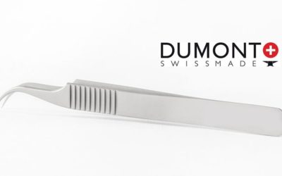
Material Metal code Tweezers Dumont
the types of materials used to make these tools, with different codes by DUMONT-Tweezers. Carbon steel Carbon steel is an extremely hard alloy (59 HRC) composed of C, Mn and Si. Although carbon provides solid spikes, it will still rust easily. This alloy is magnetic...

Safety Data Sheet : FDS or MSDS
The l Safety Data Sheet (SDS) is a form containing data on the properties of a chemical substance. The format used in North America is called MSDS (Material Safety Data Sheet). On our website, when it is necessary, you will find safety sheets, often an MSDS link that...

Codes Manufacturing materials precision tools as tweezers – forceps.
les outils de précisions sont fabriqués avec des matériaux compatibles à des applications bien définies.
On utilise des abréviations génériques pour résumé de quoi est fabriqué l’outil.
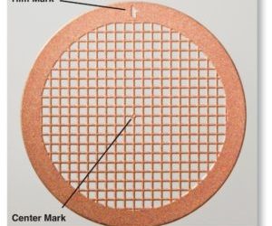
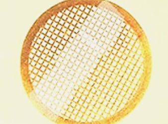
The electron microscopy grid, structure
all around the mesh of grid, the edge is commonly called rim or JANTE in French, Attention The markings materialized on the grid, can be different according to the manufacturer (Veco, Gilder, Pyser ,, ....) In the center on some grids you have a mark which makes...
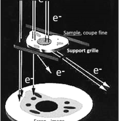
Electron Microscopy Grids: General
Electron Microscopy Grids: GeneralElectron Microscopy grids are generally used in Transmission (TEM). A TEM grid is usually a flat disk with a mesh used to support thin sections of samples such as ultra thin sections or micro-samples.The holes in the grids allow the...
fixation, E.M.?
What is the purpose of fixation for electron microscopy?"To preserve the structure of cells with minimal alteration to the living state with respect to volume, morphology and spatial relationships of organelles and macromolecules, minimal loss of tissue constituents...

E.M. grade , definition :
What does “EM grade” mean i consult a catalog of products and publications of electron microscopy, where chemicals are specified EM grade. Example: Glutaraldehyde 8%, EM grade; What does "EM-Grade" mean? Highly purified product,...

ACS grade, definition :
La désignation de qualité de réactif ACS indique la conformité aux spécifications figurant dans la dernière publication d’édition de Reagent Chemicals publiée par l’American Chemical Society…
Methods, Tips and Tricks :
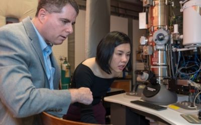
New electron microscope sees more than an image
The electron microscope, a powerful tool for science, just became even more powerful, with an improvement developed by Cornell physicists. Their electron microscope pixel array detector (EMPAD) yields not just an image, but a wealth of information about the electrons...
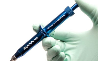
Applications of micro-punches, in sampling
A punch biopsy is a type of skin biopsy. It is a diagnostic procedure that punches a hole in the skin to acquire tissue for laboratory examination, usually through microscopy or tissue culture. Skin biopsies, compared with biopsies of other organs, are relatively...
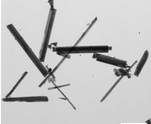
Analysis of Asbestos – Distinguishing fine fiber one by one
JEOL is industrial manufacturer of Electron Microscope and leader in this technology. has done an excellent study on the distinction between different asbestos fibers in Transmission Electron Microscopy. I would like to share with you this excellent publication.good...
My ultra-thin sections do not adhere to the grid? solutions!
Sometimes, when you try to retrieve sections on a grid, they do not stick to the surface of the grid.If you do not have time to clean the grid surfaces; Dip the grids for a moment in distilled water and remove the excess with filter paper. Let them dry; Your sections...

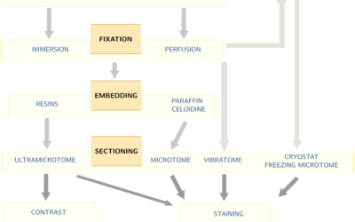
Succeed your sample preparation in Microscopy
Important parameters for your sample preparationGood sample preparation for microscopic analysis is necessary to keep tissue components as close to life as possible. For optimum conservation of biological samples, the sampling conditions are as important as the...
Electron Microscope Fixatives
Fixatives commonly used for electron microscopy- FormaldehydeCommercially available solutions are not suitable for electron microscopy because of their methanol content. Paraformaldehyde powder is used to prepare formaldehyde without methanol. The main advantage of...
UranyLess , substitute uranyle acetate
UranyLess is a new contrast for transmission electron microscopy, for your ultra-thin sections or negative stains. It may be an alternative to Uranyl Acetate, which is banned from sale due to its toxicity and problems with waste disposal.you will find a dedicated...
Microscopy Planet NEWS! :
New Microscopy Technique Shows Cells’ 3-D Ultrastructure in New Detail
The method melds the best of super-resolution fluorescence and electron microscopy LINK VIDEOoteins relate to cells’ fine structure.frame width="715" height="358" src="https://www.youtube.com/embhttps://youtu.be/HzN8d2PKI_ked/HzN8d2PKI_k" frameborder="0"...
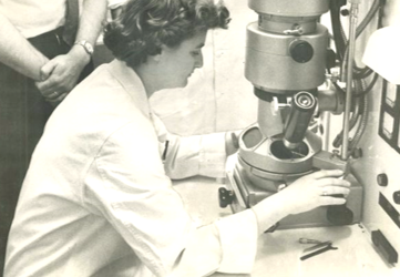
June Almeida: the woman who discovered the 1st coronavirus
The woman who discovered the first human coronavirus was the daughter of a Scottish bus driver who left school at 16.
June Almeida then became a pioneer in viral imaging, the work of which was brought to light during the current pandemic.
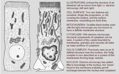
Pick from the Past Natural History, Dec. 1958, Electron Microscope
Scrutinizing the Microcosm
Electron microscopy has shown
the biologist a complex, new world.
By Huntington Sheldon.
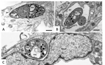
uranyless used in EM stain infection analysis for Malaria and some parasites that affect humans and animals.
An excellent work in electron microscopy using the uranyless as a contrast.In Vitro Activities of MMV Malaria Box Compounds against the Apicomplexan Parasite Neosporacaninum, the Causative Agent ofNeosporosis in AnimalsJoachim Müller 1, *, Pablo A. Winzer 2, Kirandeep...

We need a people’s cryo-EM.’ Scientists hope to bring revolutionary microscope to the masses !
this contribution by By Eric HandJan. 23, 2020 Magazine ScienceThe Laboratory for Molecular Biology (LMB), clad in glass the color of sea ice, rises like a futuristic factory above the rapeseed fields of Cambridge, U.K. It is the crown jewel of the U.K. Medical...
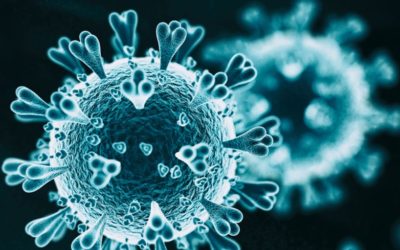
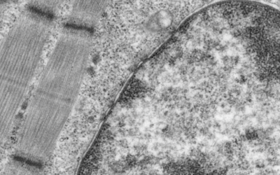
Image of the Day: Coronavirus Under the Scope
The National Institute of Allergy and Infectious Diseases releases a series of images that offer a close up look at the novel coronavirus SARS-CoV-2.source the scientist mag.
Scientists Compare Novel Coronavirus with SARS and MERS Viruses
Researchers find 380 amino acid substitutions between 2019-nCoV and severe acute respiratory syndrome (SARS)-related coronaviruses. by Abby Olena Feb 11, 2020 1.6K ABOVE: © ISTOCK.COM, KOTO_FEJAsource scientist MagAccording to a February...
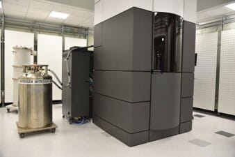
Titan Krios, the most powerful microscope in the world.
The Institut Pasteur has just launched Titan Krios, a cryogenic electron microscope which allows samples to be observed at atomic level. This cutting-edge tool will make it possible to advance knowledge on the structure of living things.To access it, you have to enter...
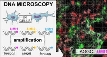
DNA microscopy
DNA microscopy makes it possible to observe both the arrangement of cells and their genetic content, thanks to an ingenious approach combining biochemistry and algorithmic reconstruction. An entirely new approach which will allow, for example, to locate mutant cells...
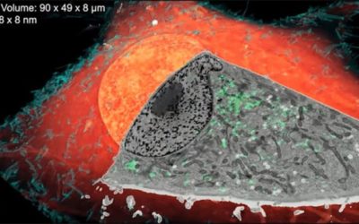
Unveiling Cells’ 3-D Ultrastructure
The method melds the best of super-resolution fluorescence and electron microscopy to show how proteins relate to cells’ fine structure. BY PRANJAL MEHAR ...
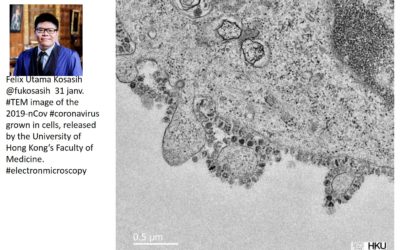
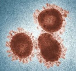
serial Killer Coronavirus
here is the first photo under an electronic microscope, the virus that makes China tremble and put on global alert: Coronavirus. the day before 2020, Dec. 31, 2019 by doctor felix Utama Kosasih Faculty of Medicine of Hong Kong,
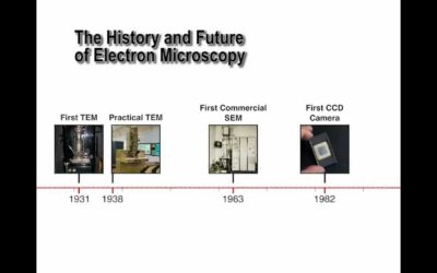
The History and Future of Electron Microscopy …. video
<iframe width="1100" height="825" src="https://www.youtube.com/embed/mx3tU5XcAlc" frameborder="0" allow="accelerometer; autoplay; encrypted-media; gyroscope; picture-in-picture" allowfullscreen></iframe> https://www.youtube.com/watch?v=mx3tU5XcAlc